V|LF-Spiro3D: respiration in images
Set up by the Multimodal Biomedical Imaging Laboratory Paris-Saclay (BioMaps - Université Paris-Saclay, CEA, CNRS, Inserm) and in collaboration with various public and private partners, the V|LF-Spiro3D project offers advances in the diagnosis of impaired respiratory function.
Asthma, pneumonia, tuberculosis, pulmonary embolism, hypertension, Covid-19... All of these diseases impair respiratory function and can lead to respiratory failure in affected individuals. To assess the severity of the impairment, pulmonologists commonly use spirometry, a pulmonary function test that measures lung function. Nevertheless, there are limitations to this test, which has prompted researchers to develop alternative diagnostic methods. The Very|Low Field 3D MR Spirometry (V|LF-Spiro3D) project, under the direction of Xavier Maître, a researcher in the BioMaps laboratory, aims to develop a new protocol for testing lung function. Using magnetic resonance imaging (MRI), the primary objective is to map lung function, in order to locate diseased areas of the lung in affected individuals. Winner of a Pathfinder 2022 grant from the European Innovation Council, awarded to projects with high innovative potential or disruptive technologies, V|LF-Spiro3D promises a veritable leap forward in the characterisation of respiratory diseases.
Spirometry: a standard that has its limits
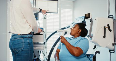
Primarily used by practitioners during pulmonary function tests for the purpose of making a diagnosis in the subjects, spirometry measures respiratory capacity via a spirometer. The protocol consists of having the subject blow through a flow meter. In a seated position, with a clip on their nose, the subject is instructed to inhale and exhale as hard as they can. The practitioner's objective is to obtain 'flow-volume' curves, in which ventilatory flows (the volume of air inhaled or exhaled by the lungs) are compared to lung volumes (the amount of air remaining in the lungs after forced exhalation). The shape of the curve indicates whether the subject has normal, restrictive or obstructive breathing.
Nevertheless, this examination has its limitations. Firstly, the breathing is forced. The subject repeatedly inhales and exhales as hard as they can. But children or people with intellectual disabilities, for example, may find it difficult to follow the instructions. Moreover, the measurement, taken at the mouth, is of both lungs together. There are therefore situations where the results do not adequately reflect the actual respiratory situation of the subject. "By drawing the line, we might not see that one of the two lungs is not functioning. Since the lung is an adaptable organ, if one of its affected regions was dysfunctional, the other regions could fully compensate for the dysfunction. The result would be a nominal flow-volume curve," explains Xavier Maître.
3D spirometry: a different approach...
The V|LF-Spiro3D project attempts to overcome these limitations by proposing a new approach: a pulmonary function test performed with an MRI machine. It envisages a ten-minute protocol in the machine. The subject lies on their back and breathes freely. The signal is obtained at this point, and the image processing provides a dynamic representation of the subject's ventilation during breathing.
The improvement is twofold: this new approach overcomes the limitations caused by the forced breathing the subject is requested to do during standard spirometry, opting instead for so-called 'spontaneous' or 'free' breathing. No particular breathing exercise is required: the subject simply lies down on the bed of the MRI machine, and the latter does the rest. The second improvement relates to the mapping of respiratory function. This mapping highlights the respiratory patterns and identifies any localised lung ventilation defects. After processing the obtained dynamic images, flow-volume curves are available for each point of the lung.
... and a technical evolution
The project has also prompted a re-think of the MRI machine. The magnetic field of standard MRI machines is usually 1.5 or 3 Tesla (T), which is 32,000 to 64,000 times greater than the Earth's magnetic field in routine clinical use. The ambition of the V|LF-Spiro3D project is to bring 3D spirometry to lower fields (0.55 T, 0.2 T or even 0.1 T). "This is lighter in terms of its economic and ecological footprint, and it wouldn't be too difficult to take this type of imaging machine out of radiology departments and bring it into pulmonary departments," explains Xavier Maître. At lower fields, differences in magnetic susceptibility at the air-tissue interface have fewer effects, which facilitates pulmonary MRI.
A European and interdisciplinary collaboration
The V|LF-Spiro3D project involves a large number of academic stakeholders (four laboratories), clinical stakeholders (four hospitals) and industrial stakeholders (Siemens and NMR Service), from four European countries (Germany, France, the Netherlands and the UK). The aim is to establish an initial database of around 500 adult and paediatric subjects, healthy and ill, covering asthma (Bicêtre Hospital, Erasmus MC in Rotterdam), chronic obstructive pulmonary disease (Pitié-Salpêtrière University Hospital), cystic fibrosis, bronchopulmonary dysplasia (Erasmus MC) and bronchiolitis obliterans after lung transplant (Foch Hospital). Together, all of these stakeholders are counting on a profound change in the characterisation of pulmonary pathophysiology and associated clinical practices. To date, the results expected with this project have no equivalent.
A patient-focused approach
Finally, the project includes an original angle of research: it questions the anthropological and philosophical dimensions behind the adoption of technology. "Does having access, not to a flow-volume curve, but to a curve 'which breathes' prompt the practitioner and the patient to take ownership of the result differently? If I see my lungs breathing, can I make anything other than a medical diagnosis?" muses Xavier Maître. By exploring the individual and cultural reaction to 3D spirometry, the project raises the question of the place of breathing in an individual's personality.
Publication :
- Boucneau T, Fernandez B, Larson P, Darrasse L, Maître X. 3D Magnetic Resonance Spirometry. Scientific Reports 10, 9649 (2020).