ReticulOme: characterising neural circuits, to act in cases of impaired motor control
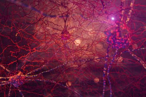
The ReticulOme project (Multi-omics characterization of descending motor circuits in the brainstem) aims to better understand the neural underpinnings of movement, with a focus on reticulospinal neurons. In the long term, it could offer a list of relevant cell types in post-traumatic or neurodegenerative contexts. The project is being conducted by the Neuronal Circuits & Motor Control Team, led by Julien Bouvier, at the Paris Saclay Institute of Neuroscience (NeuroPSI – Univ. Paris-Saclay, CNRS).
The ReticulOme project (Multi-omics characterization of descending motor circuits in the brainstem) represents the next stage in Julien Bouvier's career in the field of motricity. In 2010, Julien Bouvier, who is currently Director of Research at the CNRS, defended his thesis on the neurobiology of the respiratory system in rodents. In his thesis, the budding researcher focused on the brainstem. The following year, he left for a post-doctoral fellowship at the Karolinska Institute in Stockholm, Sweden. There his work focused on extended motricity, focusing on the role of the spinal cord and subsequently the brainstem in the control of limbs during walking, in mice. He was appointed to the CNRS in 2015, and in 2018 became head of his own research team at NeuroPSI: Neuronal circuits and motor control. In February 2023, his ReticulOme project was awarded an ERC Consolidator Grant.
A descending nerve structure
Executing coordinated movements requires complex brain processes. "These systematically involve an area of the brain known as the reticular formation. This is found in all species, because from a phylogenetic perspective it is very old. It acts as a relay for nerve signals between the brain and the spinal cord," Julien Bouvier explains. The reticular formation is located in the deepest part of the brain, the brainstem. Among other things, it houses the so-called reticulospinal neurons, which are distributed in an intertwined network in the base of the brainstem. These specific neurons come into direct descending contact with the spinal cord. This means that they are vital for the motor control of the limbs and the brainstem. "In a car accident, for example, the spinal cord can be ruptured as a result of trauma to the spinal column. This descending connectivity formed by the reticulospinal neurons is then broken, resulting in a loss of motricity which in serious cases can mean a person is unable to stand or walk", Julien Bouvier concludes.
The reticular formation, an approximate classification
Since the 1950s, many studies have focused on the reticular formation and the case of reticulospinal neurons. But the limited tools of the day only offered scientists a very broad picture of the processes at work within the nervous structure. When devices became more advanced, the reticular formation unfortunately faded into the background, with the focus now on structures involved in cognitive processes, such as the cortex. Classifying and understanding the reticular formation therefore remained at the macroscopic level, and therefore highly approximate. A situation Julien Bouvier found unfortunate: "In all likelihood, it is not a unified entity but a complex system with distinct functions. But we still lack a detailed description of the diversity of the formation at the cellular level."
The large diversity of reticulospinal neurons
However, recent work has demonstrated that there are various types of reticulospinal neurons. Each one appears to be used to control a single muscle group, in order to perform a specific type of movement. "Over the past five years, some of our work has involved isolating and characterising the functioning of one of these cell populations," Julien Bouvier reveals. To do this, his team used molecular vectors in mice, which act as a motor model for humans. The researchers then identified targeted populations of reticulospinal neurons by having them express a fluorescent protein detectable by microscopy. These same neurons were also activated or inhibited using optogenetic methods (optical and genetic techniques combined) to study the effects on the animal. At the same time, the molecular vectors followed the nerve connections by tracing the links between the neurons.
New selection criteria for reticulospinal neurons
"The main challenge of the ERC Consolidator Grant is to obtain the means to study and map all the reticulospinal neurons," Julien Bouvier stresses. And given the inherent complexity of the reticular formation, with numerous intertwined cell types, the team will have to combine multiple approaches in its research: sequencing the transcriptome (the set of genes expressed by the cell), studying the connectome (the mapping of connections), analysing cellular activity and function (summarised by the "functionome") ... Hence the suffix "Ome" in the project name! The goal of the project is to categorize reticulospinal neurons according to new criteria: anatomical (according to their position and their connection with the rest of the brain), functional (according to the type of movement they control) and genetic (according to the genes they express).
Targeting priority neural circuits
"The project is first and foremost fundamental. It aims to understand the organisation of this crucial brain structure. At the same time, it promises specific applications in the future," Julien Bouvier points out. The goal of ReticulOme is to identify priority neural circuits, with a view to motor rehabilitation therapies, by providing specialised candidates for different types of movements. Either those that facilitate walking, or, on the contrary, inhibit it, those that control posture, or those that control the movements of the brain stem or the front limbs, etc. Understanding the role played by each population, as well as their genetic identification, will serve as a starting point for designing more targeted therapies, with a view to forcing the activation or even the regrowth of neurons. And in this way, ultimately help restore a form of motricity in paralysed individuals.
Experts with multiple scientific backgrounds
To successfully implement their project, Julien Bouvier and his team will benefit from a sum of €2 million spread over five years. "We will therefore recruit two or three more people, specifically post-doctoral fellows already familiar with this field, who will enable us to develop the various experimental strands of the project. We will also enrich our technical and analytical expertise by recruiting engineers and bioinformaticians, as well as by procuring state-of-the-art equipment," predicts the Director of Research. Julien Bouvier plans to take advantage of this opportunity to highlight the overlap between specialisms, and give young researchers their place. "I hope I can be involved in training students and post-doctoral fellows on the crucial role of reticulospinal neurons in motor control. For this subject, which is still not studied enough, the idea is to create a veritable network of experts, each with his or her own scientific background," Julien Bouvier emphasises. As for the project itself, the grants should arrive by the beginning of 2024, with work starting straight away.
Publication:
- Usseglio G, Gatier E, Heuzé A, Hérent C, Bouvier J. Control of orienting movements and locomotion by projection-defined subsets of brainstem V2a neurons. Current Biology, 2020. Dec 7.
